Magnetic resonance imaging
Medical imaging technique using magnetic fields and radio waves.

UCSF Health · CC BY-SA 4.0
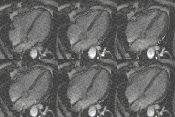
Magnetic resonance imaging (MRI) is a medical imaging technique used in radiology to generate pictures of the anatomy and the physiological processes inside the body. MRI scanners use strong magnetic fields, magnetic field gradients, and radio waves to form images of the organs in the body. MRI does not involve X-rays or the use of ionizing radiation, which distinguishes it from computed tomography (CT) and positron emission tomography (PET) scans. MRI is a medical application of nuclear magnetic resonance (NMR), which can also be used for imaging in other NMR applications, such as NMR spectroscopy.
- field
- Radiology, Medical Imaging
- known_for
- Generating detailed images of soft tissues without ionizing radiation
- key_components
- Main magnet, shim coils, gradient system, RF system
- typical_field_strength
- 1.5 T to 7 T (clinical); up to 11.7 T (research)
- contrast_mechanisms
- T1 (spin-lattice) and T2 (spin-spin) relaxation
Lore & Background
MRI was originally called NMRI (nuclear magnetic resonance imaging), but 'nuclear' was dropped to avoid negative associations. Certain atomic nuclei are able to absorb radio frequency (RF) energy when placed in an external magnetic field; the resultant evolving spin polarization can induce an RF signal in a radio frequency coil and thereby be detected. In clinical and research MRI, hydrogen atoms are most often used to generate a macroscopic polarized radiation that is detected by the antennas. Hydrogen atoms are naturally abundant in humans and other biological organisms, particularly in water and fat. For this reason, most MRI scans essentially map the location of water and fat in the body.
Reader's Guide
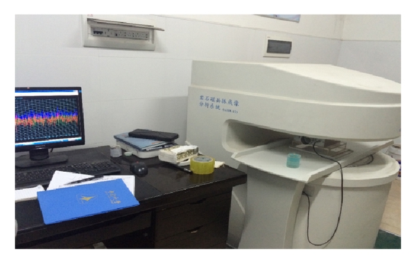
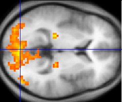
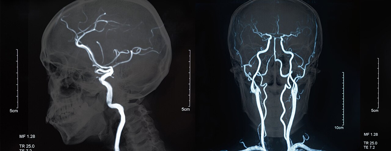
Since its development in the 1970s and 1980s, MRI has proven to be a versatile imaging technique. While MRI is most prominently used in diagnostic medicine and biomedical research, it also may be used to form images of non-living objects, such as mummies. Diffusion MRI and functional MRI extend the utility of MRI to capture neuronal tracts and blood flow respectively in the nervous system, in addition to detailed spatial images. The sustained increase in demand for MRI within health systems has led to concerns about cost effectiveness and overdiagnosis. MRI requires a magnetic field that is both strong and uniform to a few parts per million across the scan volume. The field strength of the magnet is measured in teslas – and while the majority of systems operate at 1.5 T, commercial systems are available between 0.2 and 7 T. Most clinical magnets are superconducting magnets, which require liquid helium to keep them at low temperatures. Lower field strengths can be achieved with permanent magnets, which are often used in 'open' MRI scanners for claustrophobic patients.
Did You Know?
- MRI does not involve X-rays or the use of ionizing radiation, distinguishing it from CT and PET scans.
- Hydrogen atoms in water and fat are most commonly used to generate the MRI signal.
- The characteristic repetitive noise of an MRI scan is caused by gradient coils moving slightly due to magnetostriction.
- Ultra-low field MRI (microtesla-to-millitesla range) has been demonstrated using superconducting quantum interference devices (SQUIDs).
The Physics Roots and Early Visionaries
The story of magnetic resonance imaging stretches back to the early twentieth century, when a constellation of physicists laid the groundwork that would eventually produce the diagnostic tool used in hospitals worldwide. American physicist Isidor Isaac Rabi stands at the head of this lineage; his 1944 Nobel Prize in Physics recognized his discovery of nuclear magnetic resonance, the very phenomenon that MRI exploits. In 1950, Erwin Hahn detected spin echoes and free induction decay, while Herman Carr produced a one-dimensional NMR spectrum in his 1952 Harvard doctoral thesis. Perhaps most remarkably, in 1960 a Soviet researcher named Vladislav Ivanov filed a patent for a device he called a Magnetic Resonance Imaging machine. His core insight—using magnetic field gradients paired with selective frequency excitation to encode spatial coordinates—was genuinely ahead of its time. The application was initially rejected as improbable, yet it was finally approved in 1984, preserving his original priority date. Ivanov's method, though limited to proton-density imaging and slow slice-by-slice acquisition, constituted a true imaging procedure decades before the technology became practical.
The Race to Create the First Images
The transition from spectral measurements to actual pictures of living tissue was a fiercely competitive effort spanning the early 1970s. In September 1971, Paul C. Lauterbur at Stony Brook University conceived a mechanism to encode spatial information into an NMR signal using magnetic field gradients, a theory he published in March 1973. He went on to produce the first nuclear magnetic resonance image and, in January 1974, the first cross-sectional image of a living mouse. Around the same period, Raymond Damadian, a physician at SUNY Downstate Medical Center, reported in a March 1971 Science paper that tumors could be distinguished from normal tissue in vivo by NMR relaxation differences. His 1974 patent described scanning the body to locate cancerous tissue, though it lacked a method for generating pictures. In 1976, Damadian's team captured a tumor image in a mouse thorax, and on July 3, 1977, they performed what is described as the first MRI body scan of a human being. Meanwhile, Peter Mansfield at the University of Nottingham was developing echo-planar imaging in the late 1970s, a technique that would compress scan times from hours to seconds and yield sharper images.
The Science of Tissue Contrast
Long before anyone spoke of imaging, the biophysical properties that make MRI pictures meaningful were being mapped. Physician-scientist Erik Odeblad and Gunnar Lindström described the factors governing image contrast—differences in tissue relaxation time values—nearly two decades before the first clinical scanners appeared. By 1959, Jay Singer had measured blood flow in living humans using NMR relaxation time data, and in 1967 Ligon reported NMR relaxation measurements of water in the arms of living subjects. The following year, Jackson and Langham captured the first NMR signals from a living animal, an anesthetized rat. During the 1960s, a growing body of literature addressed relaxation, diffusion, and chemical exchange of water within cells and tissues of many types. A pivotal realization arrived in the 1970s: relaxation times are the key determinants of MRI contrast and can distinguish a wide range of pathologies. Early cancer cells were found to exhibit longer relaxation times than their normal counterparts, sparking initial interest in NMR-based cancer detection by groups including Damadian, Hazlewood, and Chang. This insight also launched a program to catalog relaxation times across a broad spectrum of biological tissues, which became one of the primary motivations driving MRI's development.
From Laboratory to the Clinic
The journey from experimental curiosity to a routine diagnostic tool was marked by a handful of landmark clinical milestones. In the 1970s, a team led by John Mallard at the University of Aberdeen constructed the first full-body MRI scanner. On August 28, 1980, that machine produced the first clinically useful image of a patient's internal tissues, identifying a primary tumor in the chest, an abnormal liver, and secondary cancer in the bones. The same apparatus later served at St Bartholomew's Hospital in London from 1983 to 1993. The first clinical MRI scanners were installed in the early 1980s, and the decades that followed brought significant refinements in hardware and software, ultimately leading to the technology's widespread adoption in medicine. In 2003, Paul Lauterbur and Peter Mansfield were jointly awarded the Nobel Prize in Physiology or Medicine, recognizing their complementary contributions to the development of MRI—Lauterbur for the gradient-encoding principle and Mansfield for echo-planar imaging and related acquisition refinements. Today, MRI is a cornerstone of modern diagnostic imaging, a fact that would have seemed improbable to the researchers who first detected spin echoes in the 1950s.
Gallery
Frequently Asked Questions
Who is Magnetic resonance imaging?
MRI is a medical imaging technique in the field of radiology that produces detailed pictures of the body's internal anatomy and physiological processes. It is the clinical imaging branch of the broader nuclear magnetic resonance (NMR) family.
What are Magnetic resonance imaging's powers or role?
Its defining strength is generating high-resolution images of soft tissues without exposing the patient to any ionizing radiation. This clearly separates it from CT and PET scans, which depend on X-rays or radioactive tracers.
How does Magnetic resonance imaging work?
The scanner combines a powerful main magnet, gradient coils, and radio-frequency pulses to excite hydrogen nuclei in the body and capture their relaxation signals. Image contrast is built from T1 (spin-lattice) and T2 (spin-spin) relaxation differences between tissues.
Why is Magnetic resonance imaging important?
It gives clinicians a non-invasive window into organs, joints, and the brain that X-ray-based methods simply cannot match in soft-tissue detail. Because no ionizing radiation is involved, it can be repeated safely for longitudinal monitoring.
What are Magnetic resonance imaging's key components?
A typical clinical system includes a main superconducting magnet (usually 1.5 to 7 tesla), shim coils for field uniformity, a gradient system for spatial encoding, and an RF coil assembly for signal transmission and reception. Research-grade units can push field strengths up to 11.7 tesla.
More in Science & Measurement 1-16
Spotted an error? Know more?
This is a living reference — every entry is fact-audited, and reader corrections feed straight into our audit queue. Suggest an edit · See this site's audit record