Medical imaging
Technique for imaging body interior for clinical analysis.

Harrison Keely · CC BY 4.0
Medical imaging is the technique and process of imaging the interior of a body for clinical analysis and medical intervention, as well as visual representation of the function of some organs or tissues. It seeks to reveal internal structures hidden by the skin and bones, and to diagnose and treat disease, while also establishing a database of normal anatomy and physiology to identify abnormalities.
- field
- Medical imaging
- known_for
- Imaging interior of body for diagnosis and treatment
- inventor_of_CT
- Godfrey Hounsfield
- total_studies_by_2010
- 5 billion
- annual_chip_shipments_2015
- 46 million units
Lore & Background
The CT method is based on projecting X-rays through a section of the human head, processed by computer to reconstruct cross-sectional images.
Reader's Guide
Medical imaging has become a cornerstone of modern medicine, enabling noninvasive visualization of internal structures for diagnosis and treatment. Techniques include radiography, fluoroscopy, and magnetic resonance imaging, each with specific applications. The term 'noninvasive' applies to most imaging techniques, as no instrument is introduced into the patient's body. Interpretation is typically done by radiologists, though other healthcare professionals may also be trained.
The Spectrum of Diagnostic Modalities
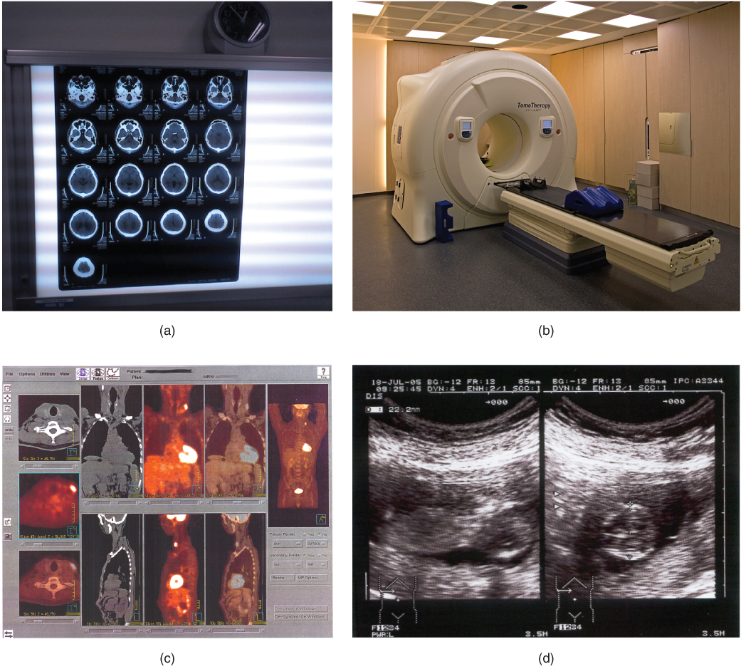
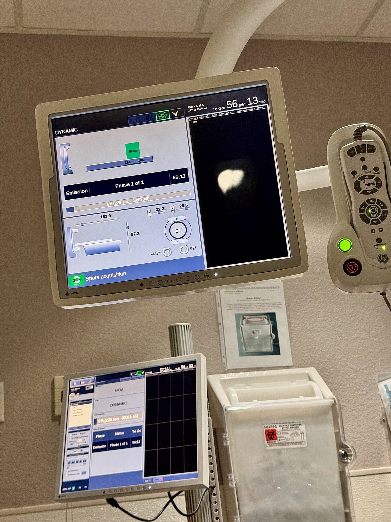
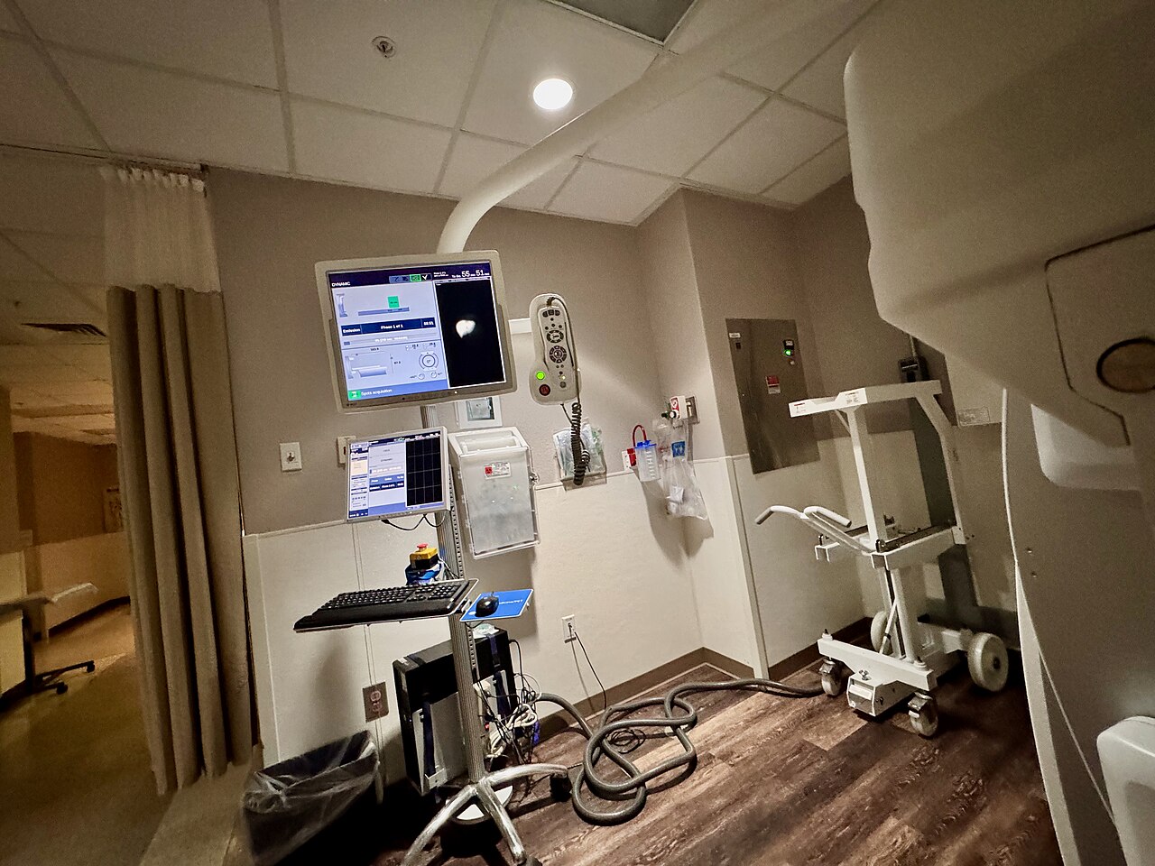
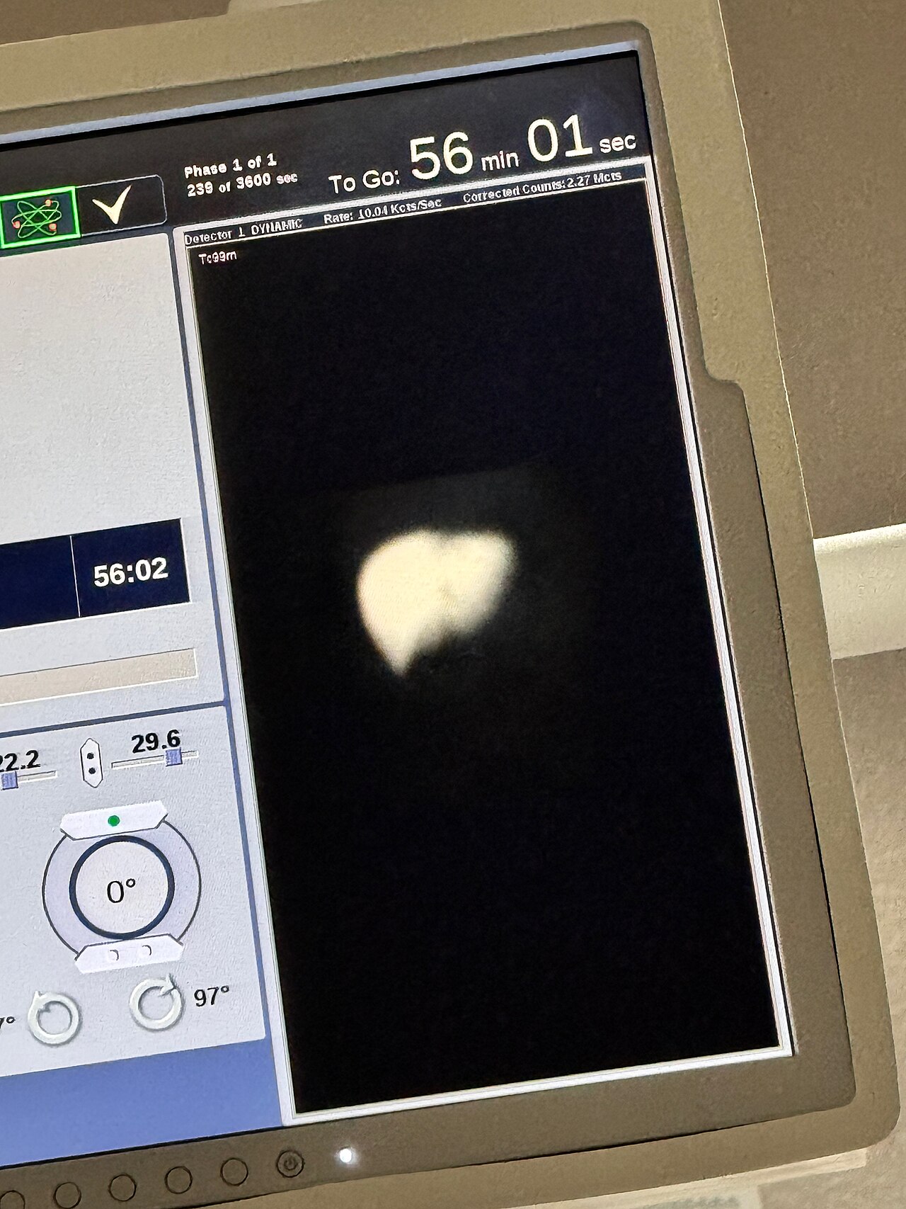
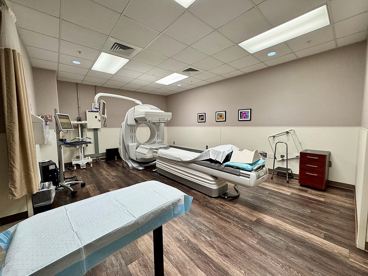
Medical imaging draws on a remarkably diverse toolkit of physical principles, each tailored to reveal something specific about the human body. At one end of the spectrum, radiography and computed tomography harness X-rays to render dense structures such as bone in sharp relief. At the other, magnetic resonance imaging taps into nuclear magnetic properties to produce detailed pictures of soft tissue entirely free of ionizing radiation. Ultrasound takes a different path altogether, firing high-frequency sound waves into the body and capturing the echoes; this makes it especially useful when real-time visualization matters, as in fetal monitoring. Nuclear medicine adds yet another dimension: positron emission tomography follows radioactive tracers through the body to expose metabolic activity rather than mere anatomy. On the frontier, photoacoustic imaging merges optical and acoustic principles into a single technique, while Magneto-acousto-electrical tomography attempts to map electrical conductivity within biological tissues. Together, these modalities ensure that no single physical phenomenon is forced to answer every clinical question.
The Imaging Chain in Clinical Practice
Every medical imaging system, regardless of modality, can be understood through a conceptual framework called the imaging chain. It starts with the patient as the subject, whose observable properties—energy emitted, reflected, or transmitted—form the raw signal. An energy source may illuminate or excite the subject to make those properties detectable. The capture device then collects that energy using the appropriate sensor: an optical system for electromagnetic radiation, a transducer for acoustic waves, or an antenna array for radio frequencies. In digital workflows, a processor takes the captured signals and converts them into a renderable format, applying algorithms for noise reduction, enhancement, or reconstruction. The final link is the display, which presents the processed information on a screen, printed film, or projection surface. Crucially, the design of every stage is informed by the characteristics of the human visual system, because the ultimate goal is to convey meaningful information to a human observer making a clinical judgment.
Coherence and the Physics of Seeing Inside
A fundamental way to classify imaging systems is by whether they use coherent or non-coherent illumination. Coherent imaging relies on an active source that produces waves maintaining a consistent phase relationship. Medical ultrasound is a prime example of this approach, as are radar, synthetic aperture radar, and optical coherence tomography. Because the phase information is preserved, coherent systems can capture not just amplitude but phase, which opens the door to sophisticated techniques like holography and interferometry. Non-coherent imaging, by contrast, depends on sources where the light waves carry random phase relationships; conventional photography, fluorescence microscopy, and telescopes all fall into this category. In the medical context, this distinction matters: it determines whether a system can exploit interference patterns to extract sub-wavelength detail or whether it must rely on intensity alone. The choice between coherent and non-coherent strategies shapes everything from the sensor design to the reconstruction algorithms that ultimately turn raw signals into a diagnostic image.
From Silver Halides to Soft Tissue
The story of medical imaging is inseparable from the broader history of photography and image capture. When Joseph Nicéphore Niépce produced the first permanent photograph through heliography in 1826, and Louis Daguerre refined the process into the daguerreotype roughly a decade later, they were establishing a scientific discipline rooted in silver halide chemistry. For much of the nineteenth century, photography remained the province of specialists. That changed when George Eastman introduced the Kodak camera in 1888 with the memorable slogan "You press the button, we do the rest," placing image-making within everyone's reach. Yet the leap into medicine came by accident: in 1895, Wilhelm Röntgen stumbled upon X-rays, a discovery that would spawn the entire field of medical imaging. World War II then proved to be a turning point, setting the stage for the rapid expansion of imaging modalities that followed in the decades after.
Gallery
Frequently Asked Questions
Who is Medical imaging?
Medical imaging is the clinical discipline of producing visual representations of a body's internal structures and organ functions. It lets practitioners see what lies beneath skin and bone without resorting to invasive procedures.
What are Medical imaging's powers or core role?
Its central ability is to reveal hidden internal anatomy, diagnose disease, and guide treatment. It also builds a reference library of normal physiology so that deviations can be flagged as abnormalities.
How does Medical imaging's story end?
There is no fixed finale; the field keeps expanding with new modalities and applications. By 2010 it had already supported roughly five billion studies worldwide, and growth has continued since.
Why is Medical imaging important to the broader science-and-measurement canon?
It is the primary non-invasive window into the human body for both diagnosis and treatment planning. Without it, clinicians would depend far more heavily on surgery or educated guesswork to identify internal problems.
Who is credited with inventing CT, and how large is the field's hardware footprint?
Godfrey Hounsfield is recognized as the inventor of computed tomography. The wider imaging hardware ecosystem is enormous—by 2015, roughly 46 million imaging-related chips were shipped each year.
More in Science & Measurement 1-16
Spotted an error? Know more?
This is a living reference — every entry is fact-audited, and reader corrections feed straight into our audit queue. Suggest an edit · See this site's audit record