
Medical ultrasound
Uses high-frequency sound waves for medical imaging and therapy.

Harrison Keely · CC BY 4.0
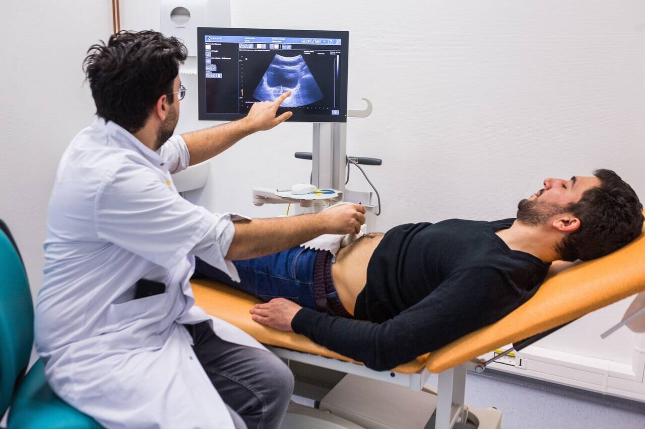
Medical ultrasound encompasses diagnostic techniques, primarily imaging, and therapeutic applications using ultrasound. It is used to create images of internal body structures such as tendons, muscles, joints, blood vessels, and internal organs, and to measure characteristics like distances and velocities. The practice of examining pregnant women using ultrasound, known as obstetric ultrasonography, was an early development of clinical ultrasonography.
- field
- Medical imaging and therapy
- types
- Diagnostic (sonography) and therapeutic
- common_modes
- A-mode, B-mode, M-mode, 3D imaging
- key_techniques
- Doppler sonography, harmonic imaging, B-flow
Lore & Background
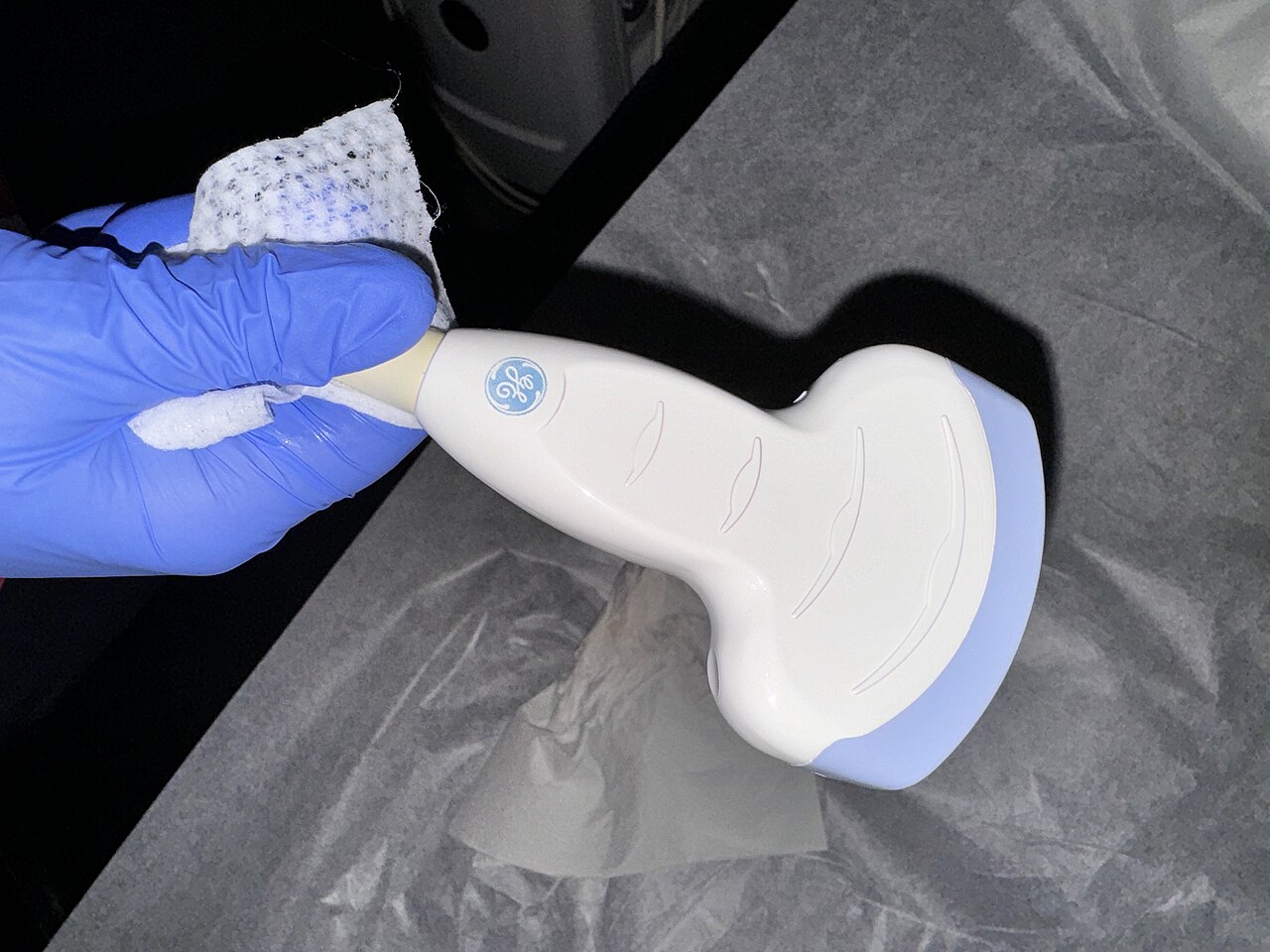
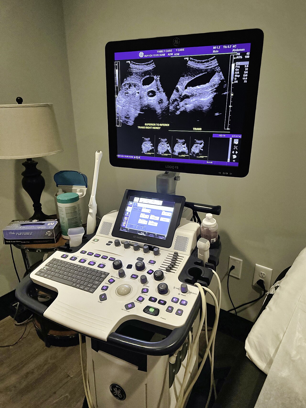
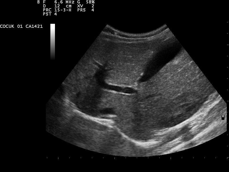
Ultrasonic images, also known as sonograms, are created by sending pulses of ultrasound into tissue using a probe. The pulses echo off tissues with different reflection properties and are returned to the probe, which records and displays them as an image. Most ultrasound examination is done using a transducer on the surface of the body, but improved visualization is often possible if a transducer is placed inside the body, using transvaginal, endorectal, or transesophageal transducers.
Reader's Guide
Medical ultrasound is significant for its wide use in diagnosis and therapy, allowing visualization of soft tissues without ionizing radiation. It is effective for imaging superficial structures at higher frequencies (7–18 MHz) and deeper structures at lower frequencies (1–6 MHz). Key applications include anesthesiology for guiding nerve blocks and vascular access, angiology for diagnosing arterial and venous disease using duplex ultrasound, cardiology via echocardiography for evaluating heart valve function and chamber size, and emergency medicine for point-of-care assessments like the FAST exam. Therapeutic ultrasound targets tumors or calculi, often using A-mode and B-mode for positioning.
Did You Know?
- A-mode is one-dimensional, while B-mode produces a two-dimensional image.
- M-mode is used to determine displacement of organ structures, most commonly the heart.
- Intravascular ultrasound uses a miniaturized probe on a catheter to image blood vessel walls.
The Physics of Sound Beyond Hearing
Medical ultrasound relies on sound waves whose frequencies exceed the roughly 20,000 Hz upper limit of human auditory perception. In a typical imaging session, a transducer—commonly called a probe—emits short pulses of these ultrasonic waves into the patient's body. As the pulses travel through tissue, they encounter boundaries between structures that have different acoustic reflection properties. The returning echoes are captured by the same probe, which then converts the electrical signals into a visual representation on a display. The machine translates the round-trip travel time of each pulse into an estimated depth, assuming a uniform sound speed of 1540 metres per second. Because real tissue varies considerably in its acoustic velocity, the resulting picture is an approximation rather than a true tomographic slice. While a general-purpose transducer suffices for most examinations, clinicians sometimes need specialized probes designed for internal placement to achieve sharper views of particular organs or vessels.
A Spectrum of Imaging Modes
Medical ultrasound offers several distinct modes, each producing a different dimensional representation of the body. In A-mode, a single transducer element fires one pulse and records the amplitude of the returning voltage as a function of two-way travel time, yielding a strictly one-dimensional trace. B-mode, or brightness mode, employs an array of elements to sweep a plane through tissue, generating a two-dimensional image in which each pixel's brightness corresponds to the backscattered signal strength. M-mode strings together successive A-mode pulses, plotting each as a bright line adjacent to the last; the result resembles a high-speed streak photograph and is especially useful for tracking the displacement of moving structures such as the heart. Three-dimensional imaging, sometimes called C-mode, stitches together multiple B-mode planes using rotating or stationary probes. A newer hybrid approach pairs ultrasound with photoacoustic excitation: a laser pulse sets hemoglobin molecules vibrating, and an arc of detectors captures the resulting acoustic signal to build a volumetric picture of blood and tissue without magnets or ionizing radiation.
Advanced Signal Techniques for Blood and Tissue
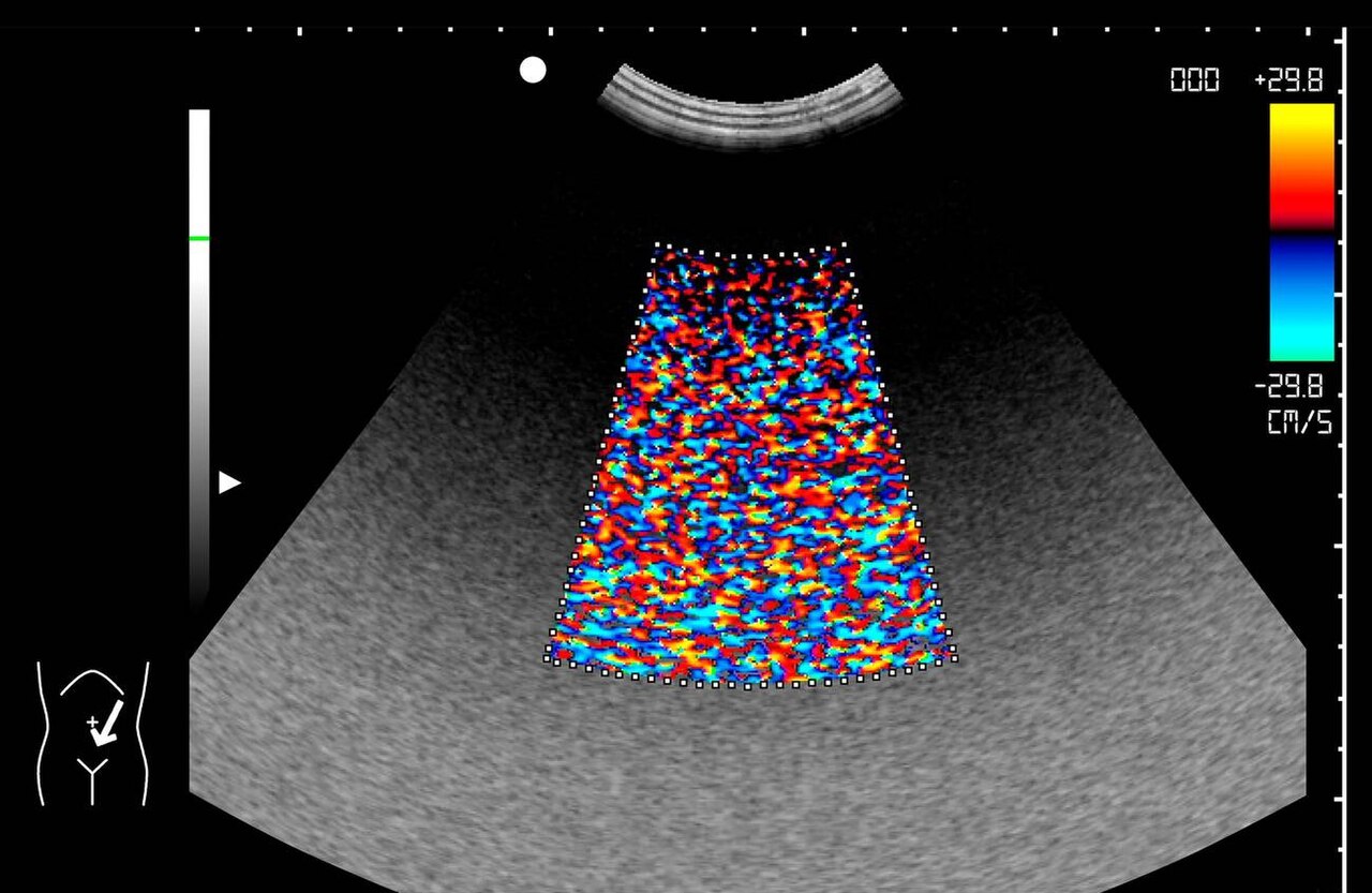
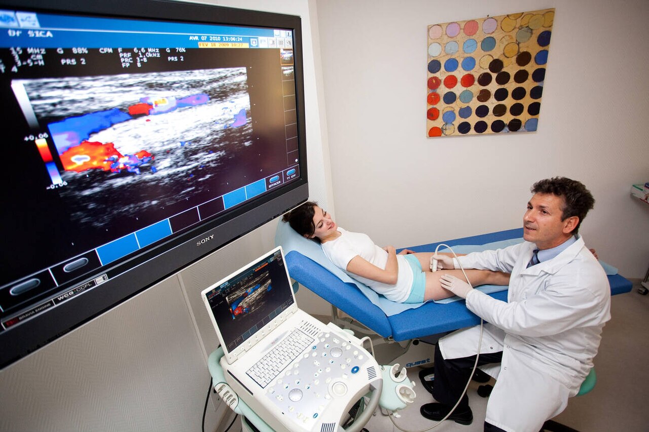
Beyond basic brightness imaging, several specialized techniques extract more information from the backscattered signal. Doppler sonography exploits the frequency shift produced when sound reflects off moving targets, most commonly red blood cells, to detect and quantify flow velocity. Harmonic imaging takes a different approach: it filters the returned signal so that only frequency components at least twice the transmitted centre frequency remain, a method particularly valuable for perfusion studies with ultrasound contrast agents and for revealing tissue-specific harmonic content. Pulse-inversion and power-modulation schemes can elicit this harmonic response without requiring real-time Fourier analysis. B-flow represents yet another strategy, digitally amplifying the weak echoes from moving blood cells while simultaneously suppressing the much stronger signals from surrounding stationary tissue, thereby rendering flowing blood and adjacent structures in a single image. It serves as an alternative or complement to conventional Doppler. When therapeutic ultrasound is directed at a tumour or a calculus, A-mode and B-mode imaging are typically employed alongside the treatment to position the probe accurately on the target region.
Clinical Roles, Terminology, and Frequency Choices
In everyday practice, sonography is a versatile tool that supports both diagnostic imaging and therapeutic guidance. Sonographers—trained medical professionals—perform the scans, and the resulting images are traditionally interpreted by radiologists or, for cardiac studies, by cardiologists. Ultrasound is particularly well suited to visualizing soft tissues, and it can guide interventional procedures such as needle biopsies or the drainage of fluid collections, making the session both diagnostic and therapeutic at once. Obstetric ultrasonography, the examination of pregnant patients, was among the earliest clinical applications of the technology. The field carries a rich vocabulary: the technique may be called sonography, echography, or ultrasonography; the equipment an ultrasound machine, sonograph, or echograph; and the output image an ultrasonogram, sonogram, or echogram. Frequency selection is a key practical decision. Superficial structures—muscle, tendon, testis, breast, thyroid, parathyroid glands, and the neonatal brain—are imaged at higher frequencies, roughly 7 to 18 MHz, because these deliver superior axial and lateral resolution, while deeper organs require lower frequencies to penetrate adequately.
Gallery
Frequently Asked Questions
Who is Medical ultrasound?
Medical ultrasound is a diagnostic and therapeutic discipline that harnesses high-frequency sound waves to visualize and treat internal body structures. It sits at the intersection of medical imaging and therapy, covering everything from sonographic imaging to therapeutic applications.
What are Medical ultrasound's powers/role?
It can produce images of tendons, muscles, joints, blood vessels, and internal organs while simultaneously measuring distances and velocities within the body. Its operational toolkit spans A-mode, B-mode, M-mode, and 3D imaging, with signature techniques like Doppler sonography, harmonic imaging, and B-flow.
How does Medical ultrasound's story end?
As a living clinical practice rather than a fixed narrative, its arc is still unfolding—obstetric ultrasonography marked one of its earliest chapters, and new therapeutic and diagnostic applications continue to be developed. The field remains an active, expanding branch of medicine with no definitive finale.
Why is Medical ultrasound important?
It offers a non-invasive, radiation-free way to examine internal anatomy and quantify physiological parameters such as blood-flow velocity. Its dual capability in both diagnosis and therapy makes it a foundational tool across many clinical specialties.
What are Medical ultrasound's signature moves?
Doppler sonography, harmonic imaging, and B-flow stand out as its most distinctive techniques, letting clinicians assess not just structure but also hemodynamic function and tissue characteristics. These methods push the practice well beyond simple anatomical imaging into functional and perfusion analysis.
More in Science & Measurement 1-16
Spotted an error? Know more?
This is a living reference — every entry is fact-audited, and reader corrections feed straight into our audit queue. Suggest an edit · See this site's audit record